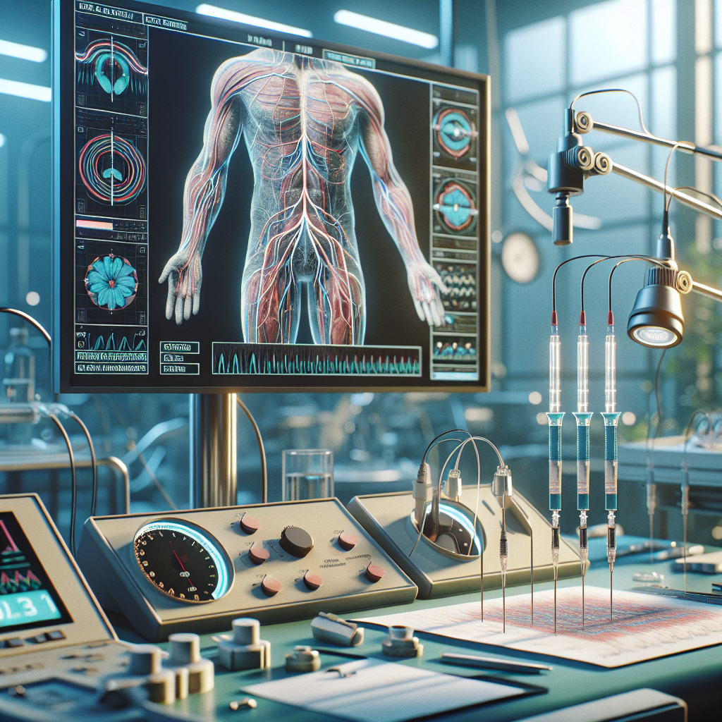
Electromyography ALS diagnosis can be a vital process that helps to identify neuromuscular disorders. This intricate, noninvasive technique monitors electrical signals in muscles, providing valuable insights into muscle and nerve function. Intense advancements in electromyography, also known as EMG, have unearthed stunning needle EMG findings and denervation patterns, particular to Amyotrophic Lateral Sclerosis (ALS).
Understanding ALS and Its Symptoms
Amyotrophic Lateral Sclerosis is a neurodegenerative condition that affects nerve cells in the brain and spinal cord, subsequently leading to muscle weakness and paralysis. The disease progression varies from person to person, but patients may experience a gradual onset of muscle weakness or stiffness, difficulty swallowing, and even loss of ability to move or speak in advanced stages.
It is important to note, ALS is not always easy to diagnose because its symptoms can be similar to other neurological disorders. Hence, the role of tools like electromyography ALS diagnostics becomes crucial.
Significance of Electromyography ALS Diagnosis
One of the primary tests used in the diagnosis of ALS is electromyography. An EMG is a diagnostic procedure that evaluates the health condition of muscles and nerve cells that control them. It leverages tiny needle-like electrodes called needle EMG which are inserted into a muscle to record electrical activity.
Stunning Needle EMG Findings in ALS
The needle EMG findings in ALS are remarkable and include a variety of patterns. Firstly, patients with ALS will often show signs of active denervation. This means there is an ongoing loss of nerve supply to particular muscles.
The second interesting pattern discernible in many ALS patients is that of chronic reinnervation, which is the body’s compensation mechanism by forming new nerve-muscle connections where other connections have been lost. ALS patients may show increased motor unit potential amplitude, duration, and complexity, indicating ongoing muscle remodeling due to chronic reinnervation.
A third needle EMG finding that is striking in ALS is widespread denervation. EMG testing can often demonstrate that denervation is taking place in a variety of places throughout the body, not just in the area initially affected, reflecting the systemic nature of this disease.
Decoding Denervation Patterns in ALS
In the broader context of the disease, the study of denervation patterns in ALS provides us with more precise and valuable information. Recent studies have shown that denervation in ALS often starts focally before spreading widely, allowing us to realize that early diagnosis and intervention can potentially slow the disease progression.
Studies performed using needle EMG have shown that denervation and reinnervation can also provide insight into prognosis. Patients whose muscles show a higher rate of reinnervation often fare better than those in whom reinnervation is less successful.
Final Thoughts
In conclusion, the role of electromyography ALS diagnostics and the study of needle EMG findings and denervation patterns provides a solid foundation for understanding this complex disease. Armed with these sophisticated diagnostic tools, medical professionals are better equipped to diagnose and manage this challenging condition.
If you or a loved one are grappling with ALS or have had exposure to harmful stimuli like Real Water, remember you are not alone. The community at lasvegasalsrealwater.com extends its support and services for individuals coping with ALS.
For specialized assistance, don’t hesitate to reach out through our website’s contact page or explore more related content on our blog page. For immediate assistance, feel free to call us at 702-385-6000. We are here to assist you every step of the way.
References:
– Mayo Clinic – Amyotrophic lateral sclerosis (ALS)
– National Institute of Neurological Disorders and Stroke – Amyotrophic Lateral Sclerosis (ALS) Fact Sheet
– PubMed – Diagnostic criteria for amyotrophic lateral sclerosis
– The New England Journal of Medicine – Amyotrophic Lateral Sclerosis
– Johns Hopkins Medicine – Electromyography (EMG)