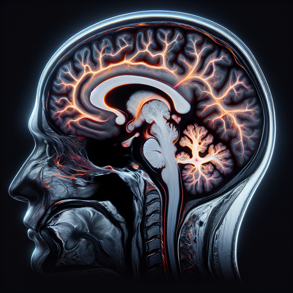
Brain MRI ALS (Amyotrophic Lateral Sclerosis) has emerged as an indispensable tool in the medical community for diagnosing this devastating illness while simultaneously ruling out other diseases. Accurate diagnosis of ALS, a progressive neurodegenerative disease that affects nerve cells in the brain and spinal cord, can be challenging due to the similarity of its symptoms to other neurological disorders. As such, brain MRI and spinal cord imaging play a crucial role in providing detailed images of the nervous system, helping doctors rule out other potential conditions and decrease uncertainty in ALS diagnosis.
Importance of Brain MRI in ALS Diagnosis
The diagnosis of ALS typically involves ruling out other potential conditions exhibiting similar symptoms— a process often termed as a ‘diagnosis of exclusion.’ Here, brain MRI ALS comes into play as a credible, non-invasive tool.
MRI, or Magnetic Resonance Imaging, produces detailed images of soft tissues in the body, including the brain and spinal cord. These images may reveal signs of diseases like Multiple Sclerosis, tumors, stroke, or other brain irregularities, which can cause symptoms akin to ALS. If the brain MRI does not reveal these-related signs, the physician can consider ALS diagnosis.
Spinal Cord Imaging and Its Role in ALS Diagnosis
Just as crucial is the role of spinal cord imaging. While MRI of the brain may not always show changes specific to ALS, the combination of brain and spinal cord imaging can provide a robust analysis and aid in a more confident ALS diagnosis.
Various imaging techniques utilized for spinal cord can identify certain signature traits of ALS. Detailed spinal cord imaging may reveal atrophy or thinning of the spinal cord, particularly in the neck region or cervical spinal cord. Such changes, although not exclusively, can correlate with ALS, providing further diagnostic clarity.
Ruling Out Other Diseases with ALS Brain MRI and Spinal Cord Imaging
Physicians utilize brain MRI ALS and spinal cord imaging to rule out other ailments manifesting similar symptoms to ALS. These diseases could range from spinal cord tumors, cervical spondylosis, neurosarcoidosis, and spinal vascular malformations, to mimic conditions like multifocal motor neuropathy or spinal muscular atrophy.
Moreover, brain and spinal cord imaging can reveal the presence of other neurodegenerative diseases such as Multiple Sclerosis that could coexist with ALS. Accurate differentiation and diagnosis is key to formulating an effective treatment plan.
Advancements and Future Potential
Studies are currently underway to identify MRI biomarkers that show changes exclusive to ALS. Future breakthroughs in this direction could significantly contribute to streamlining ALS diagnosis process.
Rest assured, the evolving field of brain MRI ALS and spinal cord imaging holds the potential to revolutionize the diagnostic approach to ALS. Armed with this state-of-the-art technology, medical professionals can expedite accurate diagnosis, plan targeted treatments, and facilitate better patient outcomes.
References
– Amyotrophic Lateral Sclerosis (ALS) Fact Sheet
– MRI in the diagnosis and management of ALS
– Update on MRI in amyotrophic lateral sclerosis
Looking to learn more about your specific ALS case and how Brain MRI ALS might be instrumental in your circumstance? We invite you to reach out through our contact page on the lasvegasalsrealwater.com website. Our expert team is on standby to assist you. Want additional related content? Navigate to our blog for insightful articles on ALS and associated aspects. Alternatively, for immediate assist, do not hesitate to call us at 702-385-6000. Your journey towards understanding ALS doesn’t have to be arduous; we are here to help.